Dental bone augmentation forms the biological foundation for long-term restorative and implant success. However, experiencing a failed dental bone graft can cause significant distress and delay planned smile restoration. While bone grafting procedures boast high clinical success rates, surgical complications, micro-movements, or bacterial ingress can sometimes compromise healing.
Recognising complications early allows oral surgeons to intervene promptly. Timely assessment helps salvage surrounding jaw architecture and prevent irreversible tissue loss. Whether you recently underwent ridge preservation or sinus augmentation, understanding the warning signs of a failed bone graft is essential for safeguarding your oral health and future implant plans.
Failed Dental Bone Graft Complications and Biological Integration
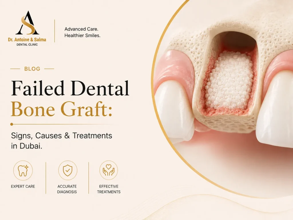
A dental bone graft acts as a cellular scaffold to stimulate new bone regeneration. Over several months, your body replaces the graft material with vital native bone through osteoconduction and osteogenesis. A failed dental bone graft occurs when this biological fusion is interrupted, causing the material to resorb prematurely, loosen, or develop an active bacterial infection.
When bone fails to integrate around an existing site or adjacent tooth, the structural base becomes compromised. Identifying a failed bone graft tooth site requires distinguishing normal post-surgical tenderness from pathological failure.
- Normal Post-Surgical Healing: Mild swelling peaking at 48 hours, light pinkish oozing on day one, and dull tenderness manageable with prescribed pain relief.
- Pathological Graft Failure: Progressively worsening pain, persistent tissue redness, loose granules shedding after the first week, and visible purulent drainage.
Addressing these issues requires structured clinical evaluation using high-resolution 3D imaging to assess remaining graft density.
Recognising Critical Failed Dental Bone Graft Signs
Early detection plays a pivotal role in saving compromised tissue, making close monitoring essential during the initial recovery window. While moderate discomfort and swelling should subside within three to four days, intense, pulsating pain radiating to the jaw, neck, or ear signals acute inflammation or potential graft rejection. Similarly, swelling that worsens past the third postoperative day or presents with shiny, dark red gum tissue indicates that the site is struggling to heal.
Signs of active infection and structural compromise demand immediate clinical attention. Any visible yellow or green discharge oozing from the incision margins—often accompanied by a foul taste and persistent halitosis—points to an infected bone graft. Additionally, while minor granule shedding during the first 24 hours is normal, losing substantial amounts of loose bone material or experiencing gum flap separation that exposes the underlying membrane leaves the graft vulnerable to oral bacteria.
Patients must also remain vigilant regarding unresolved soft tissue numbness or tingling that extends past the expected anaesthetic period, as this may indicate compression or irritation of adjacent sensory nerves. Recognizing these combined warning signs early allows for prompt intervention to manage infection, alleviate nerve pressure, and preserve the integrity of the graft.
Comparison of Normal Post-Surgical Healing vs. Failed Dental Bone Graft
| Clinical Parameter | Normal Post-Operative Healing | Failed Dental Bone-Graft Complications |
| Pain Level & Duration | Mild to moderate; steadily decreases after 48–72 hours | Intense, throbbing, or worsening past day 4 |
| Swelling & Gum Colour | Peaks at 48 hours, then recedes; pink, healthy tissues | Progressively increasing swelling; dark red, shiny margins |
| Graft Material Shedding | Few stray granules within first 24 hours only | Continual washing out of granules past the first week |
| Fluid / Drainage | Light pinkish serous fluid on day 1 | Yellow/green purulent discharge (pus) with foul taste |
| Incision Line (Flap) | Sutures remain intact; margins closed securely | Flap reopening (dehiscence) exposing bone/membrane |
| Systemic Symptoms | None, normal body temperature | Possible fever, fatigue, or swollen lymph nodes |
Primary Causes Behind A Failed Bone Graft
A successful graft relies on pristine sterile technique, robust blood supply, mechanical stability, and meticulous patient aftercare. When a failed dental bone graft occurs, it is generally linked to specific clinical or physiological factors.
1. Bacterial Contamination and Infection
Oral bacteria can infiltrate the surgical pocket during the procedure or throughout the postoperative phase. An untreated infected bone graft in mouth tissue destroys the osteogenic matrix before new blood vessels can form.
2. Insufficient Vascularisation and Blood Supply
Bone cells require oxygen and systemic nutrients delivered through microscopic blood vessels. Compromised blood flow—often exacerbated by systemic conditions or tight soft-tissue tension—prevents cellular infiltration.
3. Premature Mechanical Disturbance and Pressure
Micromovement disrupts delicate capillary networks forming inside the graft. Chewing hard foods directly on the surgical area, vigorous mouth rinsing, or tongue probing can dislodge the bone matrix.
4. Patient-Specific Health and Lifestyle Factors
- Tobacco and Vaping Use: Nicotine acts as a potent vasoconstrictor, drastically reducing peripheral blood circulation and elevating failure rates.
- Uncontrolled Systemic Conditions: Poorly regulated diabetes or autoimmune conditions impair standard inflammatory response cycles.
- Poor Plaque Control: Inadequate oral hygiene allows harmful oral biofilms to build up along delicate suture lines.
Corrective Failed Dental Bone Graft Treatment In Dubai
Experiencing graft failure does not mean dental implants are out of reach. Advanced restorative protocols allow specialists to debride compromised sites and rebuild volume safely. Accessing targeted failed dental bone graft treatment in Dubai provides patients with advanced surgical intervention tailored to complex anatomy.
1. Advanced 3D CBCT Diagnostic Evaluation
Specialists utilize low-dose Cone Beam Computed Tomography (CBCT) to evaluate the exact boundary of bone loss, bone density changes, and sinus floor stability.
2. Surgical Debridement and Decontamination
Under profound local anaesthesia or conscious sedation, the surgeon reopens the site to remove non-integrated granules, infected granulation tissue, and compromised membranes. The underlying bony bed is thoroughly irrigated with antimicrobial solutions.
3. Site Sterilisation and Soft Tissue Management
Before placing any revision material, the anatomical area must be entirely free from active pathogen colonies. Specialists often allow the soft tissue to heal for several weeks to ensure a vascular, infection-free bed.
4. Secondary Bone Augmentation
Once the oral environment stabilizes, a secondary graft is placed using premium biomaterials, autologous growth factors (PRF/PRP), and high-grade barrier membranes to guarantee superior stability.
Essential Measures for Preventing Graft Complications
Preventing a failed bone graft tooth complication begins with stringent surgical protocols and disciplined home care routines.
- Strict Adherence to Prescription Protocols: Complete the full course of prescribed antibacterial rinses and oral antibiotics without early discontinuation.
- Adopting a Soft-Food Diet: Consume nutrient-rich, lukewarm, and soft foods for the first two weeks to avoid physical pressure on the surgical site.
- Total Nicotine Cessation: Refrain entirely from smoking, vaping, or shisha use for at least two weeks before and several weeks after surgery.
- Gentle Oral Hygiene: Clean surrounding natural teeth carefully while avoiding direct mechanical brushing over the healing graft margins.
- Choosing a Specialist-Led Surgical Centre: Ensure your treatment is executed by licensed oral surgeons or periodontists experienced in advanced regenerative dentistry.
Specialist Oral Rehabilitation Care at A&S Clinic
Managing complex bone revisions requires clinical precision, sterile surgical environments, and bespoke restorative planning. At A&S Clinic Dubai, our surgical team combines extensive diagnostic expertise with cutting-edge regenerative techniques to address compromised grafts and restore long-term oral stability.
Located on Al Wasl Road in Al Manara, our clinic offers comprehensive dental implantology, bone augmentation, and periodontal revision therapies in a comfortable setting. Every treatment plan is customized to protect your dental health and deliver predictable, lasting aesthetic results.
Restore Your Oral Health with Precision Care at A&S Clinic
If you suspect complications with your dental graft, prompt evaluation is crucial to protecting your smile.
Contact the surgical team at A&S Clinic in Dubai today to schedule your comprehensive diagnostic consultation and secure your pathway to healthy dental restoration.
Conclusion
Navigating the recovery of a dental bone augmentation requires vigilance, as timely detection is the key to salvaging compromised jaw structures. While encountering a failed dental bone graft can feel like a setback, modern oral surgery provides highly effective, predictable revision protocols to restore bone density and stability.
Identifying issues like persistent swelling, pain, or an infected bone graft in mouth tissues early allows specialists to intervene before irreversible bone loss occurs. With accurate 3D diagnostics, sterile surgical debridement, and tailored regenerative care, your path toward a permanent dental implant remains completely achievable.
Frequently Asked Questions
What are the earliest failed dental bone graft signs?
The earliest signs include persistent throbbing pain beyond four days, worsening gum swelling, loose bone granules continually emerging from the site, and visible pus discharge.
Can an infected bone graft in mouth tissue be saved?
Mild superficial infections may resolve with targeted antibiotics and clinical irrigation, but deeply compromised, non-integrated grafts typically require surgical removal and subsequent re-grafting.
How soon can a failed bone graft be redone?
Following thorough cleaning and removal of failed material, surgeons generally allow soft tissues to heal for eight to twelve weeks before performing a revision graft.
Does smoking increase the risk of a failed dental bone graft?
Yes. Nicotine restricts localized blood flow and impairs immune response, significantly increasing the likelihood of poor bone integration and bacterial infection.
Is revision failed dental bone graft treatment painful?
No. Revision procedures are conducted under modern local anaesthesia or conscious sedation, ensuring complete intraoperative comfort alongside tailored postoperative pain management.
How does a dentist confirm a failed bone graft?
Dentists evaluate clinical symptoms alongside 3D CBCT scans and digital radiographs to check for lack of bone density, spatial movement, or structural bone loss.